The early detection of oncological pathology has become a priority for the Romanian state. Thus, in order to support patients and facilitate access to diagnosis, a new medical investigation dedicated to oncological suspicion has been introduced, with unlimited reimbursement.
Atlas Imagistica - Computer Tomography
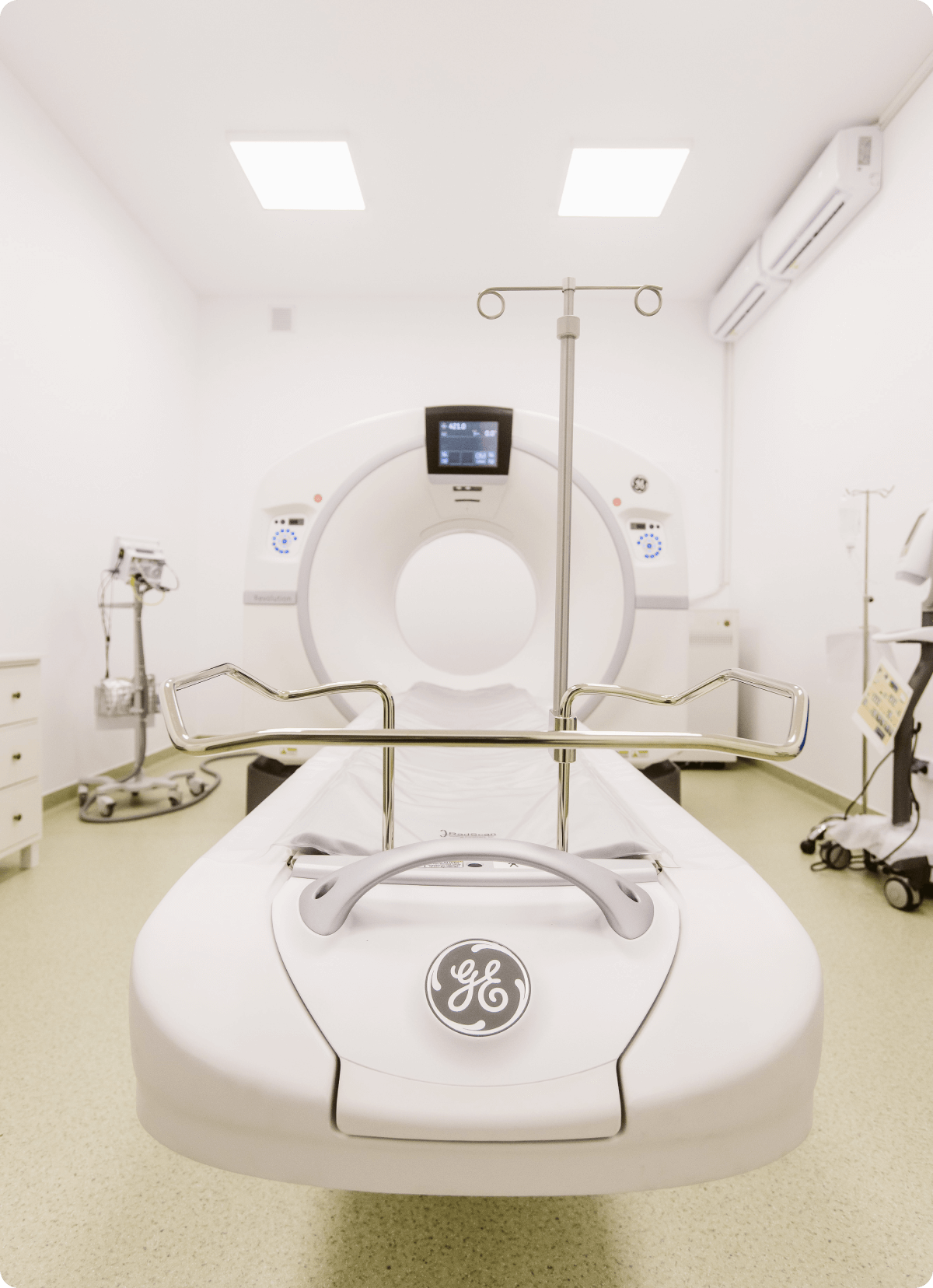
What is Computer Tomography?
Computer Tomography (CT) is a useful diagnostic tool for examining structures inside the body. A CT scan uses X-rays and computers to produce images of a cross-section of your body. It creates pictures that show very thin 'slices' of bones, muscles, organs, and blood vessels.
The rapid acquisition of CT data means that precise images and information can be obtained without being influenced by patient movement, whether physiological (e.g., cardiac or respiratory). CT scans can be performed with or without contrast material, which can be administered orally or injected into a vein, thereby providing additional details of organs.

Preparing for CT
Before presenting yourself for a CT scan, it is important to ensure that a CT scan is the best test to confirm or exclude the suspected condition. If you are unsure, contact a radiologist and ask for advice, especially before requesting a CT scan in a child or young person, due to their higher radiosensitivity. Other techniques may be more suitable for these patients.
It is important to exclude early pregnancy in a fertile-aged woman, as this is usually an absolute contraindication for CT.
We recommend performing serum creatinine to assess kidney function, if the situation requires the administration of contrast material.
Contraindications
Individuals with a history of allergy to iodine-based contrast material
Pregnant women
Chronic kidney failure
What is Cardiac CT Angiography?
It is an increasingly common medical imaging examination. This is primarily due to its excellent negative predictive value in excluding coronary artery disease. However, the image resolution and, therefore, the diagnostic outcome are limited by the patient's heart rate and rhythm. Adequate patient preparation is essential to achieve a diagnostic result.

Preparing for Cardiac CT Angiography
Take all usual medications on the day of the examination, especially blood pressure medications, except for oral antidiabetic drugs that should be stopped on the day of the examination and for an additional 24/48 hours after the investigation.
It is recommended not to consume caffeine-containing products on the day of the examination, as they could hinder efforts to reduce heart rate before the examination. These products include: coffee, tea, energy drinks, energy pills, and weight loss pills.
For CT angiography, serum creatinine measurement is necessary. Patients should also consume fluids before and after the administration of the contrast material for renal protection.
A target heart rate for CT angiography is 65 bpm or less. Beta-blocker medications are considered frontline for achieving short-term heart rate reduction. These medications can be used orally, intravenously, or both. The most common approach uses oral metoprolol with doses based on the resting heart rate, ranging from 25 mg to 50 mg, administered 30 minutes to 1 hour before scanning.
If premedication is considered safe (consultation with a cardiologist), then a possible regimen would be to administer 50 mg of oral metoprolol 12 hours before the scan and an additional 25-50 mg of oral metoprolol 30 minutes/1 hour before the examination.
Indications for Cardiac CT Angiography
Noninvasive assessment of coronary artery anomalies and other thoracic vessels
Symptomatic patients with low/moderate probability of coronary artery disease
Normal or uninterpretable ECG
Normal or equivocal cardiac biomarkers
Non-acute symptomatic patients with moderate risk and no known heart conditions
Non-acute symptomatic patients with low risk and no known heart disease (if the patient cannot exercise or undergo a stress test)
Assessment of newly occurring heart failure
Newly occurring heart failure
No history of coronary artery disease, low/intermediate probability
Reduced ejection fraction
Preoperative evaluation of coronary arteries before non-coronary cardiac surgery
Discordant ECG
New or worsening symptoms with normal previous stress imaging studies
Preoperative assessment for transcatheter aortic valve implantation (TAVI/TAVR) of a coronary bypass graft
What Regions Can We Evaluate with CT?
Cerebral – evaluates the anatomy of the cerebral parenchyma, used in headaches, dizziness, evaluation of the vascular system in ischemic vascular accidents, bleeding due to trauma or aneurysm ruptures, and in brain tumors
Neck (cervical region) – we can evaluate inflammatory or infectious processes, neoplasms in the pharynx, larynx, and esophagus; response and monitoring of oncological therapy, vascular malformations, thyroid pathology, congenital anomalies
Spine – for evaluating fractures, congenital anomalies, vertebral static changes (scoliosis), detecting tumors or metastases from various organ types
Cardiac – cardiac or coronary anatomy, to detect or diagnose coronary artery disease, to assess the permeability of coronary bypass grafts or implanted coronary stents, or to evaluate cardiac volume and function (including ejection fraction)
Thorax – used for detecting pulmonary infections, tumor formations (pulmonary, mediastinal, pleural), pleural or pericardial fluid, respiratory tract, rib cage, and vascular anomalies
Abdomen and Pelvis – infections such as appendicitis, pyelonephritis, or infected fluid collections (abscesses), inflammatory bowel diseases (ulcerative colitis or Crohn's disease), pancreatitis or liver cirrhosis, tumors in various organs (liver, kidney, pancreas, ovary, bladder, intestine), calculi (biliary, renal, bladder), aneurysms (aortic, iliac, renal), and monitoring the treatment response of oncology patients
Angiography - evaluating the vascular system throughout the body, to detect atheromatous plaques, degree of stenosis, aneurysms, and vascular malformations
Musculoskeletal System - for detecting fractures, tumor formations, and inflammatory joint changes


