ATLAS IMAGISTICA
Centre of Excellence in Medical Imaging | Targu-Mures, Romania
Blog Article | Series: Prevention & Patients | October 2026 - Breast Cancer Awareness Month
A screening mammogram takes a few minutes. Waiting for the result should not take weeks.
Why breast cancer screening by mammography saves lives, and why the time to result is part of the quality of care
October, Breast Cancer Awareness Month: a simple step with measurable impact
Breast cancer is the most common cancer in women in Romania. In 2022, an estimated 12,685 new cases and 3,877 deaths were recorded. Five-year survival is 75%, compared with the European Union average of 83%, and the difference is attributed mainly to low participation in screening.
A study conducted in 2025 across four centres in Bucharest, on 2,500 women diagnosed with breast cancer, showed that 76% had never had a mammogram before diagnosis. October, dedicated to breast cancer awareness, is the right moment to restate a simple fact: regular mammography is the most effective method of early detection.
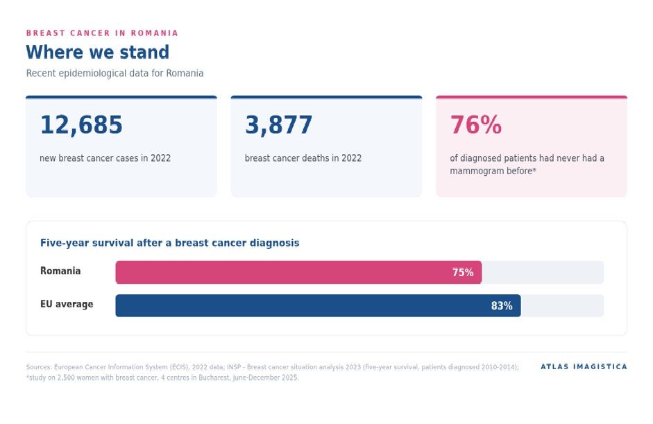
Figure 1. Breast cancer in Romania - recent epidemiological data.
Why mammographic screening makes the difference
The prognosis of breast cancer depends to a large extent on the extent of the disease at the time of diagnosis. When the tumour is confined to the breast, relative five-year survival exceeds 99%. When the disease has spread to the lymph nodes, it falls to 87%, and in the presence of distant metastases, to approximately 33%.
Mammography can identify lesions several years before they become palpable. Organised programmes in Romania address women aged 50 to 69, with a mammogram every 2 years, while European guidelines extend the recommended range to 45-74 years. In 2026, free regional programmes were launched, such as the DARIA project coordinated by the Cluj-Napoca Oncology Institute, which targets 119,000 women. The decision on the starting age and the interval is taken together with the family doctor or the specialist, according to individual risk.
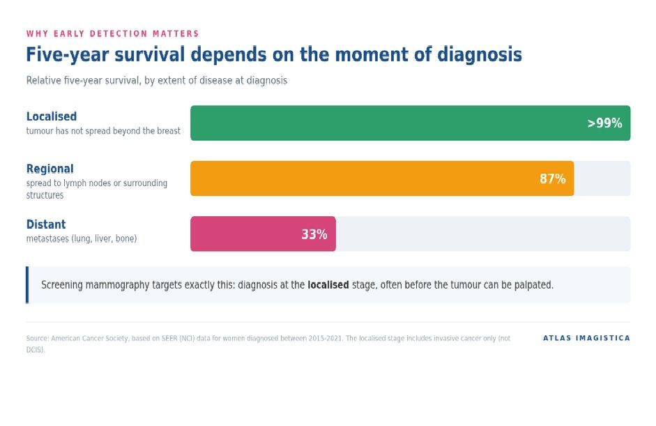
Figure 2. Relative five-year survival in breast cancer, by stage at diagnosis.
THE PERSPECTIVE OF PHYSICIANS AND CLINICS
What makes the difference in a mammographic interpretation
The value of a mammographic screening programme depends on two indicators that pull in opposite directions: the cancer detection rate and the recall rate. A good interpretation workflow balances them. Three practices, recommended by European and international guidelines, have the greatest impact.
Double reading of mammograms
The European Commission guidelines for breast cancer screening and diagnosis (ECIBC) recommend, in organised mammographic screening programmes, double reading of mammograms, with discordances resolved by consensus or arbitration. Double reading identifies more cancers than reading by a single physician, while consensus or arbitration keeps the number of recalls under control.
Within the Atlas network, screening mammograms can be interpreted independently by two radiologists, and in case of discordance a third physician decides.
Comparison with previous mammograms
Comparison with previous mammograms makes it possible to distinguish stable changes from new or evolving ones. It is one of the most effective factors in reducing recalls for known benign findings and, by extension, unnecessary anxiety.
The Atlas radiologist has access within the platform to the patient's previous examinations performed in the network, and the relevant imaging history (date and BI-RADS category of previous examinations) is recorded in the report.
Structured BI-RADS reporting
The ACR BI-RADS system (5th edition) standardises the vocabulary, the structure of the report and the assessment categories. For the referring physician, a structured report means a clear conclusion and explicit management; for the clinic, it means reports that are comparable over time and measurable quality indicators. A structured mammography report contains:
- the type of examination and the views performed (for example CC and MLO, with or without tomosynthesis);
- breast density, in ACR categories a-d;
- description of the lesions according to the BI-RADS lexicon: location (quadrant, distance from the nipple), size, shape, margins, associated calcifications;
- comparison with previous examinations;
- the final BI-RADS assessment category (0-6) and the management recommendation.
Atlas mammography reports follow this structure and include the BI-RADS category and the management recommendation in the conclusion.
How we shorten the interval from mammogram to report at Atlas Imagistica
The Atlas Imagistica teleradiology network is built to reduce the interval between the mammographic examination and the report, without compromising the quality of interpretation:
- Secure transfer. The mammogram performed at the partner clinic arrives through the TeleradiologyNET platform, encrypted, within approximately 10 minutes of the examination being completed.
- Automatic allocation. In under 2 minutes, the case is assigned to an available radiologist with experience in breast imaging, also taking into account the same patient's previous examinations.
- Comparison with previous examinations. Comparison with previous mammograms reduces unnecessary recalls and, by extension, interval ②.
- 24/7 delivery. The report reaches the teleradiology portal and the partner clinic's PACS. The referring physician can discuss the case directly with the radiologist.
Median reporting time for mammography within the Atlas network: 24.4 working hours (from registration of the study in the Atlas system to signature of the report; 119 mammograms, October 2025 - September 2026; excluding weekends and public holidays).
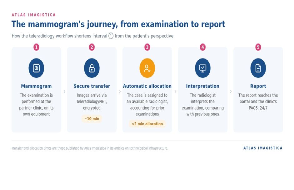
Figure 5. The screening mammography workflow within the Atlas Imagistica teleradiology network.
For clinics and hospitals
Running mammographic screening campaigns? The Atlas Imagistica teleradiology network provides mammogram interpretation 24/7, with the report delivered directly into your systems.
Madalina Vîrtopeanu
Business Development and Partnerships Manager
+40 724 551 580 | [email protected]
31 Mihai Viteazul Street, Targu Mures
[email protected] | +40 770 341 500 | atlasimagistica.com
THE PATIENT'S PERSPECTIVE
„For the patient, the examination is a brief moment. The interval until the result is the part that is felt most. A sound imaging workflow treats this interval as part of the medical act.”
The longest part of a mammogram: waiting for the result
The specialist literature is consistent: a mammogram with a normal result does not produce lasting anxiety. Anxiety arises in the intervals of uncertainty, particularly when the patient is recalled for further investigation.
- In a prospective study, 46% of recalled women had borderline or clinically significant anxiety before the recall visit. Anxiety decreased significantly within a few days in those informed of a benign result.
- Women with a false-positive result received prescriptions for anxiolytics or antidepressants 10-20% more frequently than those with an immediate negative result.
- Patients perceive the period of investigation as longer than the one recorded in the hospital's documents.
- 97% of women stated that receiving the result immediately would reduce their anxiety.
- A false-positive result increases the likelihood that some women will not return for the next screening round. The National Cancer Institute recommends, as a solution, clarifying abnormal results on the same day.
The time to result therefore influences not only the patient's comfort, but also long-term participation in screening.
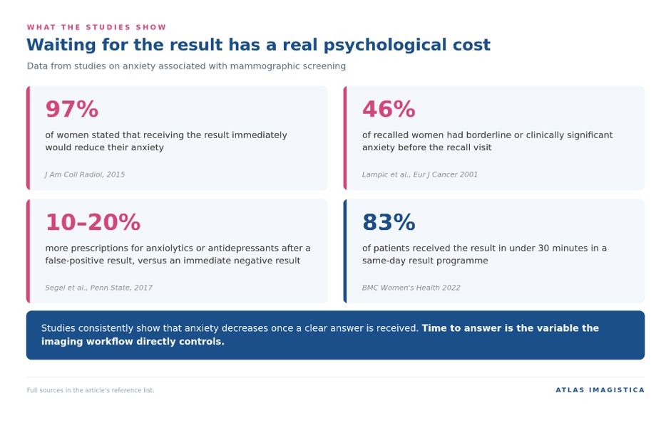
Figure 3. Anxiety associated with waiting for the mammogram result - data from studies.
The three waiting intervals after a mammogram
From the patient's perspective, the path from mammogram to a clear diagnosis may include three distinct intervals:
- ① Mammogram → report. The interval between the examination and receiving the result. It is the only interval every patient goes through, and it depends directly on the imaging interpretation workflow.
- ② Recall → further investigation. Additional views, tomosynthesis or ultrasound. Most recalled women do not have cancer: in a Norwegian study of 526 recalled women, 87.6% ultimately had a false-positive result.
- ③ Biopsy → histopathology result. The interval associated with the highest level of stress, and one that depends on the pathology laboratory.
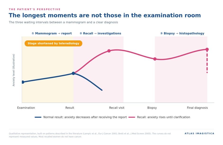
Figure 4. The three waiting intervals after a mammogram and the qualitative course of anxiety.
How to read your mammogram result: the BI-RADS categories explained
The mammogram result is expressed as a BI-RADS category, the standardised classification system used in mammographic reporting. Knowing what each category means reduces uncertainty after receiving the report.
Category
Meaning
What follows
BI-RADS 0
Incomplete assessment
Additional views, ultrasound or comparison with previous examinations are required. It does not mean a diagnosis of cancer.
BI-RADS 1
Negative
Routine screening, at the recommended interval.
BI-RADS 2
Benign findings
Routine screening. The findings described (cysts, benign calcifications, known fibroadenomas) require no further investigation.
BI-RADS 3
Probably benign (malignancy risk ≤2%)
Short-interval imaging follow-up, usually at 6 months.
BI-RADS 4
Suspicious
Biopsy recommended.
BI-RADS 5
Highly suggestive of malignancy (≥95%)
Biopsy and prompt referral to the oncology team.
BI-RADS 6
Biopsy-proven malignancy
Examination performed in the context of treatment planning.
Practical recommendations for the day of the mammogram
- Bring your previous mammograms and reports. Comparison reduces the likelihood of a recall.
- Do not apply deodorant, talc or cream to the underarm and breast area on the day of the examination; they can create false images.
- If you are premenopausal, schedule the examination in the week after your period, when the breasts are less tender.
- Ask when and how you will receive the result. A known timeframe reduces uncertainty.
- If you are recalled, schedule the further investigations as soon as possible. In most cases, the final result is benign.
References
- European Cancer Information System (ECIS). Breast cancer incidence and mortality estimates, Romania, 2022.
- National Institute of Public Health. Situation analysis - breast cancer, 2023.
- Study on mammographic history in 2,500 women with breast cancer, 4 centres in Bucharest, June-December 2025 (communicated by the Romanian Society of Breast Surgery).
- American Cancer Society. Breast Cancer Survival Rates (SEER data, women diagnosed 2015-2021).
- „Prof. Dr. Ion Chiricuță” Oncology Institute, Cluj-Napoca. The DARIA project - free mammographic screening for 119,000 women, 2026-2029.
- Brett J, et al. The psychological impact of mammographic screening. A systematic review. J Med Screen, 2005.
- Lampic C, et al. Short- and long-term anxiety and depression in women recalled after breast cancer screening. Eur J Cancer, 2001.
- Direct interactive public education by breast radiologists about screening mammography: impact on anxiety and empowerment. J Am Coll Radiol, 2015.
- Segel et al. False-positive mammogram results and anxiety/depression medication use. Penn State University, 2017.
- Impact of same day screening mammogram results on women's satisfaction and overall breast cancer screening experience. BMC Women's Health, 2022.
- National Cancer Institute. Some women avoid breast cancer screening after false-positive mammogram results. Cancer Currents, 2024.
- Recall mammography and psychological distress. Norwegian study of 526 recalled women (PubMed 23021930).
- European Commission Initiative on Breast Cancer (ECIBC). European guidelines on breast cancer screening and diagnosis - the recommendation on double reading. Joint Research Centre, European Commission.
- American College of Radiology. ACR BI-RADS® Atlas, 5th edition, 2013.
About Atlas Imagistica
Atlas Imagistica is a Centre of Excellence in Medical Imaging based in Targu-Mures, Romania, founded and led by Dr. Sorin Roșca MD. We provide AI-assisted teleradiology services to hospitals and clinics in Romania 24 hours a day, 365 days a year.